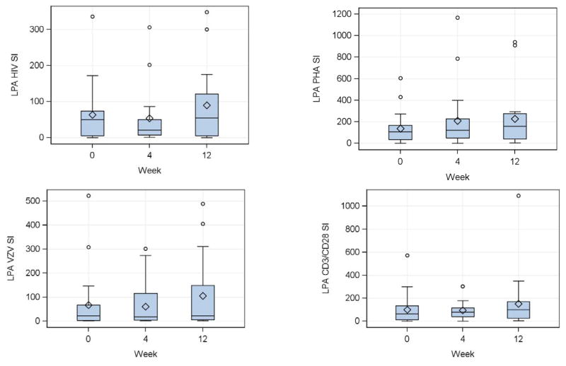
Figure 1. Effect of a single dose of DMPA on lymphocyte proliferative responses in HIV-infected women.
Data were derived from 24 HIV-infected women. Proliferation was measured by 3H-Thymidine incorporation after 6 days of in vitro stimulation of PBMC collected at the indicated time points. Stimulation indices (SI) calculated by the median incorporation in stimulated PBMC divided by the median incorporation in unstimulated or mock-stimulated controls, for HIV antigen (upper left), VZV antigen (lower left), PHA mitogen (upper right) and CD3/CD28 ligands (lower right) are summarized in box plots showing the medians as horizontal lines inside the boxes; means as diamonds inside the boxes; upper and lower quartiles as the box boundaries; minimum and maximum values, excluding potential outliers, as whiskers and outliers as open circles. There were no significant changes from week 0 (DMPA administration) to week 4 (peak MPA concentration) or week 12 (maximum exposure to MPA).

