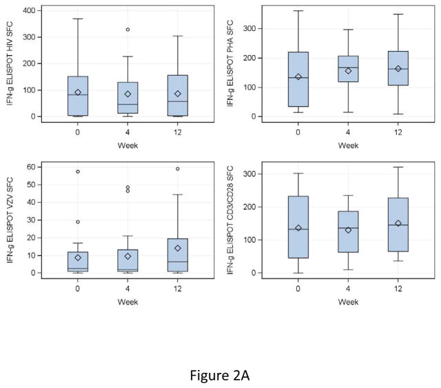
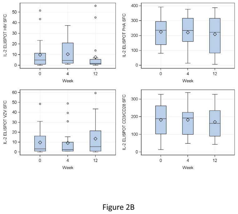
Figure 2. Effect of a single dose of DMPA on ELISPOT responses in HIV-infected women.
Data were derived from 24 HIV-infected women. Interferon γ (IFNg; Panel A) and IL2 secretion (Panel B) were measured by dual-color fluorospot. Adjusted spot forming cells (SFC)/105 PBMC calculated by subtraction of the mean SFC in control unstimulated or mock-stimulated wells from the mean SFC in stimulated PBMC, for HIV antigen (upper left), VZV antigen (lower left), PHA mitogen (upper right) and CD3/CD28 ligands (lower right) are summarized in box plots showing the medians as horizontal lines inside the boxes; means as losanges inside the boxes; upper and lower quartiles as the box boundaries; minimum and maximum values, excluding potential outliers, as whiskers and outliers as open circles. There were significant increases from week 0 (DMPA administration) to week 12 (maximum exposure to MPA) in VZV IFNγ and IL2 SFC (p=0.007 for both). SFC in other conditions remained unchanged.


