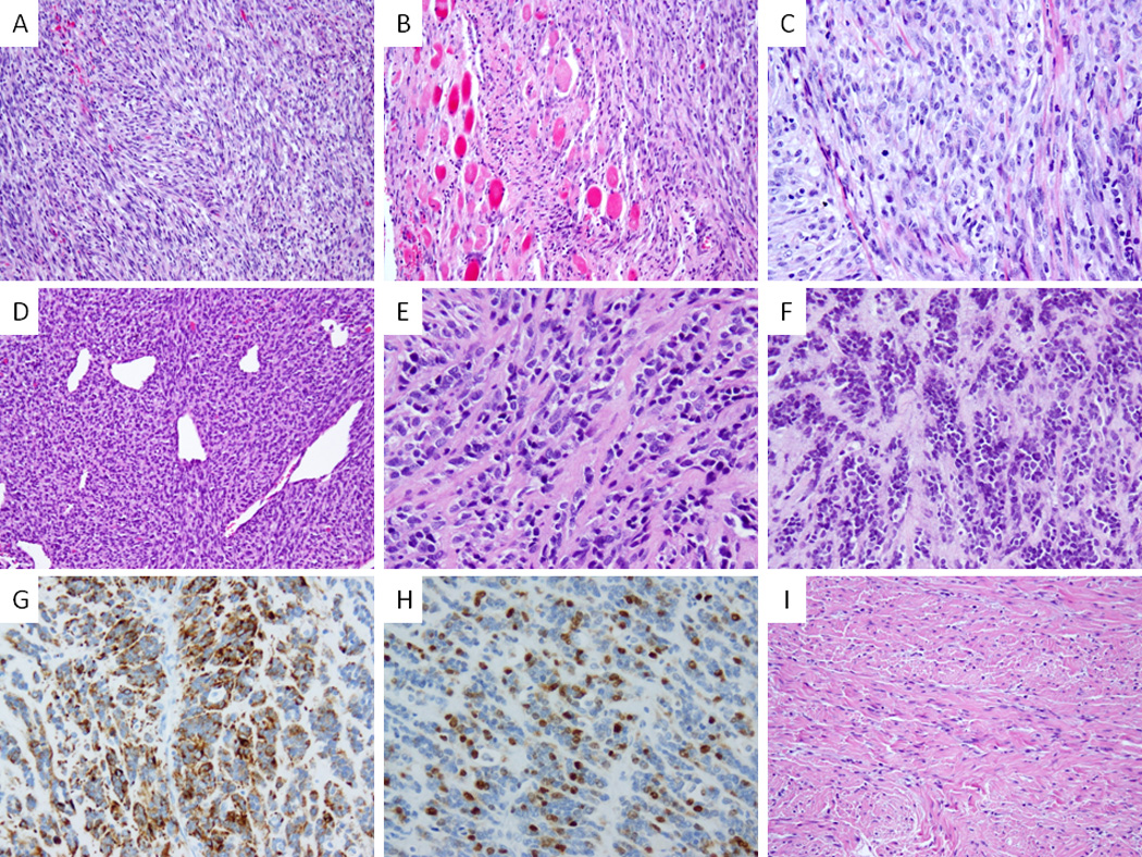
Figure 3. Pathologic features of congenital/infantile fusion positive SRMS.

VGLL2-CITED2 fusion positive tumors shared similar morphology (A–C) with monomorphic spindle cells arranged in short intersecting fascicles (A, SRMS6), infiltrating within skeletal muscle (B, SRMS6) and showing a more plump ovale cells with pale eosinophilic to clear cytoplasm, fine chromatin and scattered mitotic figures (C, SRMS7). VGLL2-NCOA2 fusion positive tumor (SRMS3) showed a highly cellular and hyperchromatic appearance reminiscent of infantile fibrosarcoma with a distinctive hemangiopericytoma-like vascular pattern in the primary tumor (D); in the subsequent two local recurrences, 2 years (E) and 5 years (F) later, it had a more sclerosing appearance; latter recurrence showed reactivity for desmin (G) and myogenin (H). In contrast a VGLL2 rearranged tumor with no identifiable partner showed a more sclerotic background mimicking fibromatosis (SRMS10, I).
