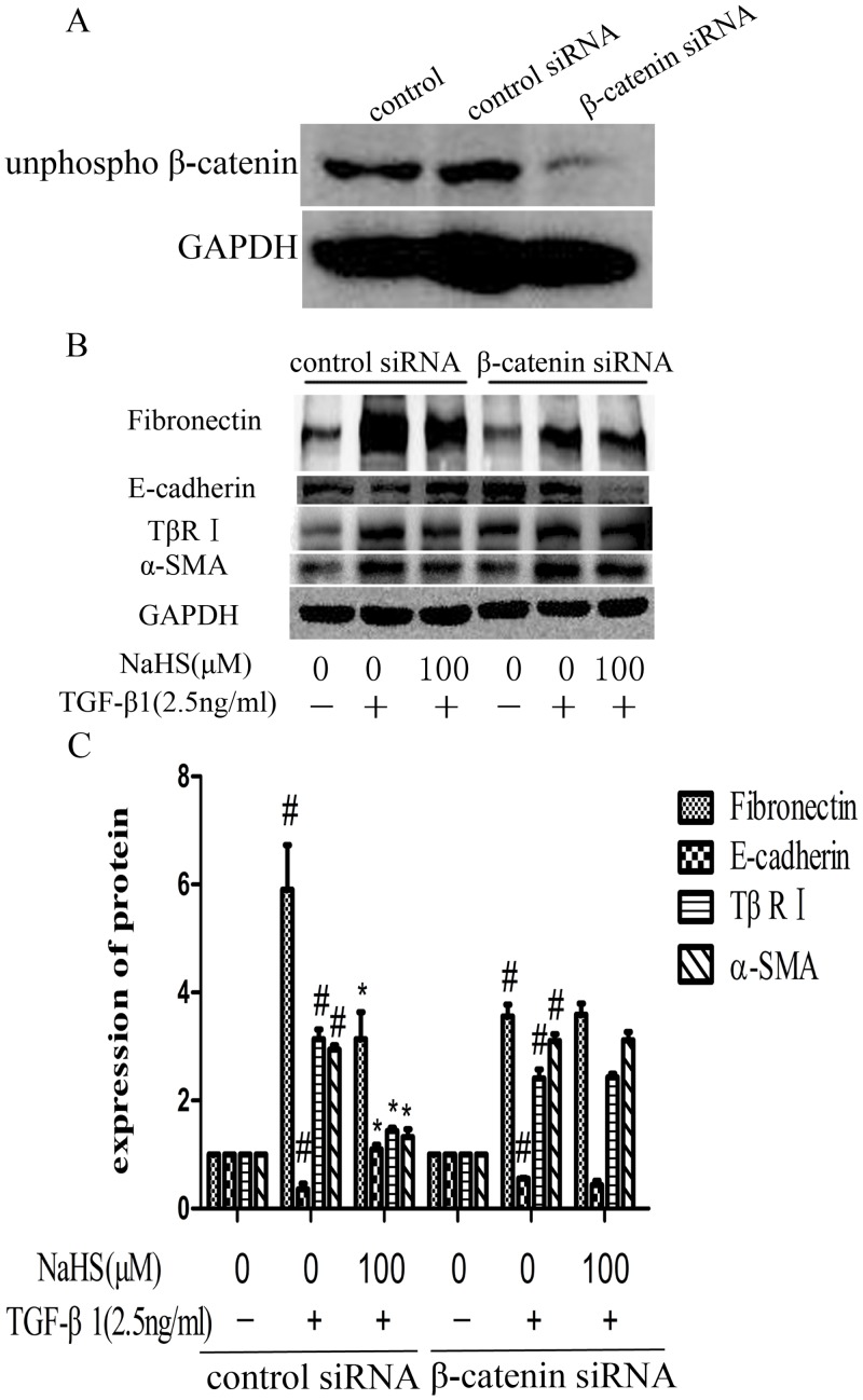
Fig 8. NaHS attenuates the TGF-β1 induced EMT via Wnt/catenin pathway.
(A) β-catenin knockdown efficiency by siRNA in HK-2 cells. The cells were transfected with β-catenin siRNA or control siRNA for 6 hours and the culture medium was replaced by fresh medium. Western blot assay was used to detect the expression level of non-phosphorylated β-catenin. (B) After transfected with β-catenin siRNA or control siRNA for 6 hours, HK-2 cells were incubated with NaHS (100μM) for 12 hours and then with TGF-β1 for the following 36 hours. The expression level of fibronectin, E-cadherin, TβR I and α-SMA were examined by western blot assay. (C) Graphical representation of the relative quantification for the protein level. The relative values were calculated by the density of targeted proteins vs GAPDH (%). The values of mean ± SEM (n = 3) were gained from relative abundance quantified by densitometry and normalized to GAPDH. #P<0.05 vs. control group, *P<0.05 vs TGF-β1 group. One-way ANOVA followed by Tukey’s multiple comparison test.

