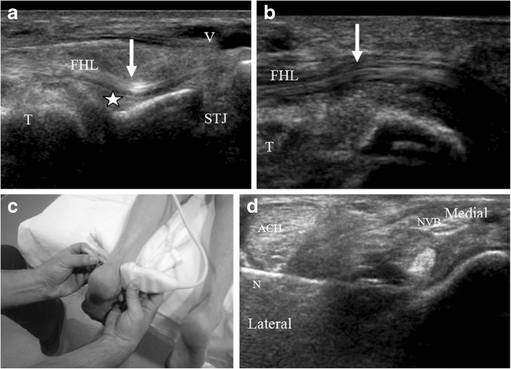
Fig. 2.
a and b Longitudinal images below the level of the medial malleolus demonstrates kinking (arrow in a) of the flexor hallucis longus (FHL) tendon proximal to the subtalar joint (STJ) from scarring of the posterior recess of the ankle joint (asterisk), reflecting posterior impingement. b Post FHL tendon sheath release surgery longitudinal imaging at the same level demonstrates mass effect on the FHL tendon to have resolved (arrow in b) (T talus). c, d Ultrasound showing FHL peritendinous injection using a lateral peri- Achilles (ACH Achilles tendon) approach in prone position with probe placed on medial skin side. This approach allows access to the FHL with the needle (N) while avoiding the posterior tibial neurovascular bundle (NVB). e, f Cine images before and after surgery similarly demonstrate smoother motion of the FHL tendon after surgical release with active plantar and dorsiflexion of the big toe (ESM 8, ESM 9).

