Fig. 1.
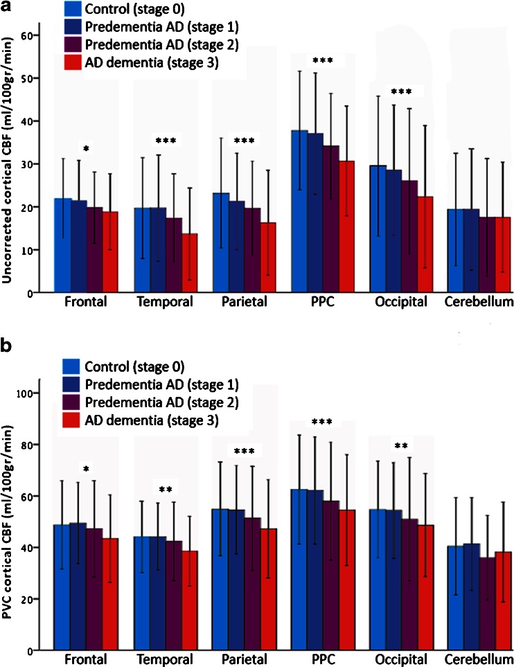
Lower regional cerebral blood flow (CBF) is associated with advancing Alzheimer’s disease (AD) stage. Bar graphs display mean regional uncorrected CBF (a) and mean partial volume corrected (PVC) cortical CBF (b) for the different AD stages, per brain region. Error bars represent standard deviations (±2SD). Dose-response relationships between CBF and AD stage were detected using general-linear models (AD stage entered as a continuous measure), resulting in a p for trend < 0.05 in all brain regions except the cerebellum. The most prominent relationships between decreasing CBF and advancing AD stage were found in the posterior supratentorial brain regions (p for trend < 0.001). PPC precuneus and posterior cingulate cortex. *p for trend < 0.05, **p for trend < 0.01, ***p for trend < 0.001

