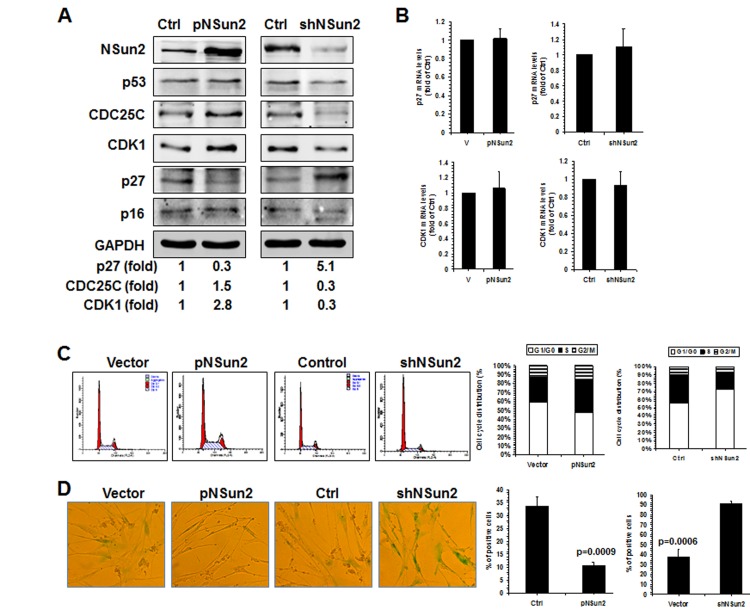
Figure 5. NSun2-p27 regulatory process impacts on the progression of replicative senescence.
(A) 2BS cells were stably infected with a lentivirus bearing a pHBLV-NSun2 vector (pNSun2) or a pHBLV-shNSun2 vector (shNSun2). Cell lysates were prepared and subjected to Western blot analysis to assess the levels of proteins NSun2, p53, CDC25C, p27, p16, CDK1, and GAPDH. (B) RNA prepared from cells described in Fig. 5A was subjected to RT-qPCR analysis to assess the levels of p27 and CDK1 mRNAs. Data represent the means ± SD from 3 independent experiments. (C) Cells described in Fig. 5A were analyzed for cell cycle distribution (left). The percentage of cells in each cell cycle compartment is presented (right). (D) Cells described in Fig. 5A were analyzed for the activity of SA-β-gal (left). The means ± SD from 3 independent experiments are presented; significance was analyzed by Student's t test (right).

