Figure 9.

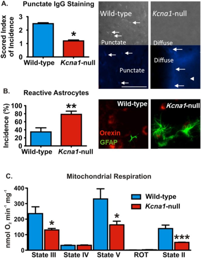
Pathology is apparent in the lateral hypothalamus/perifornical region (LH/P) of Kcna1-null mice. (A) Photomicrographs depict immunoglobulin G (IgG) labeling (white/blue) in LH/P parenchyma. In wild-type animals, punctate staining (arrows) is more pronounced, compared to diffuse labeling in Kcna1-null mice. Bar graph of punctate mouse IgG staining as scored on an arbitrary 1–5 scale by five blinded investigators (n = 4 animals/group, 3–4 LH/P sections/animal). (B) Reactive astrocytes are GFAP-positive (green) and exhibit extended processes and hypertrophy (see the Kcna1-null darkfield photomicrograph) when compared to typical astrocytes as depicted in the wild-type section. Coronal sections containing LH/P had a significantly higher incidence of reactive astrocytes compared to wild-type LH/P (n = 4 animals/group, 3–4 LH/P sections/animal). Sections are double-labeled for orexin (red). (C) Hypothalamic mitochondria isolated from Kcna1-null mice have reduced state III, state V, and state II respiratory rates when compared to wild-type controls (n = 3–4). Data are mean ± standard error of the mean. *P < 0.05, **P < 0.01, ***P < 0.001.
