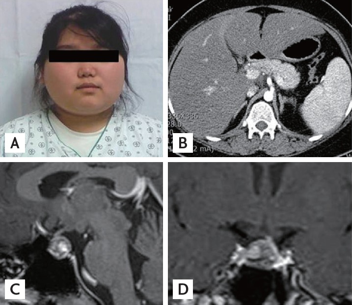
Figure 1.

(A) Round and plethoric face of the patient. (B) Abdomen computed tomography of the patient showing normal adrenal glands. (C, D) Sellar magnetic resonance imaging of the patient showing a 1.5-cm, markedly heterogeneous pituitary macroadenoma with left-sided stalk deviation.
