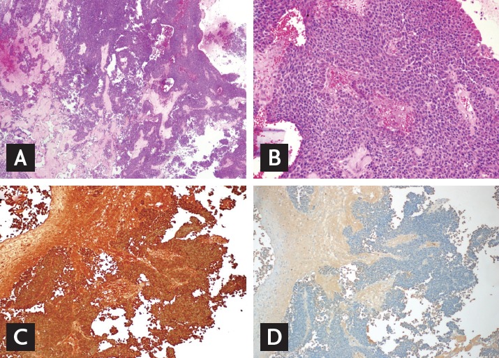
Figure 3.

Tissue pathology of the pituitary tumor. The tumor cells were arranged in a trabecular and diffuse pattern (A: H&E, ×40). They were with granular eosinophilic cytoplasm, occasionally forming focal pseudovascular rosettes (B: H&E, ×200). On immunohistochemistry, the tumor was positive for adrenocorticotropic hormone (C: ACTH, ×100), but growth hormone (D: GH, ×100), prolactin, thyroid stimulating hormone, follicle stimulating hormone, and luteinizing hormone were all negative (data not shown). These pictures were provided by courtesy of Dr. Se Hoon Kim in Yonsei University.
