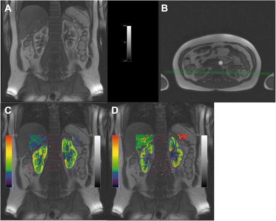
Fig. 9.

Example using a data set with different slice orientation and UMMperfusion. a and b two slices from the DCE-MRI acquired following the protocol described in [57], comprising four coronal slices (a) and one transversal slice (b). c map of the plasma flow calculated by UMMPerfusion and the 2CFM superimposed on (a, d) map of the extraction fraction derived superimposed on (a), respectively
