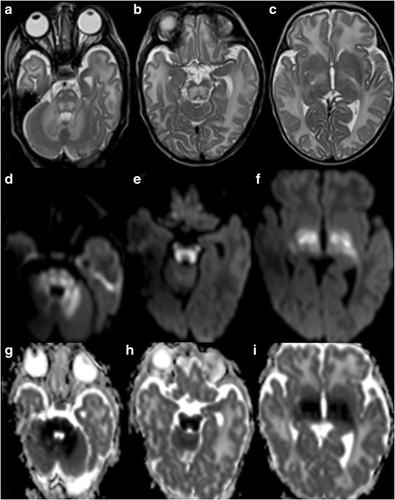
Fig. 2.

a-c, Axial T2-weighted MR images of a 20-day-old male term neonate with maple syrup urine disease show swelling and hyperintense signal of the cerebellar white matter, dorsal pons, corticospinal tracts along its course in the basis pontis, midbrain, and posterior limbs of the internal capsule, and thalami. d-f, Trace of diffusion images and g-i, Apparent diffusion coefficient (ADC) maps of the same newborn reveal bright DWI-signal and matching low ADC values, respectively, in the cerebellar white matter, dorsal pons, corticospinal tracts in the basis pontis, midbrain, and posterior limb of the internal capsule, and thalami representing restricted diffusion/cytotoxic edema compatible with extensive ongoing injury to the myelinated parts of the brain
