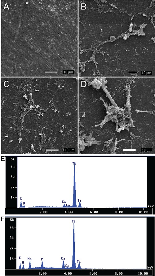
Figure 6. Scanning electron microscopy and energy dispersive X-ray spectroscopy analyses of titanium disks. Scanning electron microscopy revealed that untreated titanium disks (A) exhibited few and small particle deposits. Titanium disks coated with sodium hyaluronate (HY; B), single-walled carbon nanotubes (SWCNT; C), and HY-functionalized SWCNT (HY-SWCNT; D) showed higher particle deposition compared with uncoated disks. Energy dispersive X-ray spectroscopy analysis showed that the deposited particles in untreated titanium disks contained only calcium ions (E), while in disks treated with SWCNT, HY or HY-SWCNT (F), the particles contained calcium, sodium, and phosphorus ions.

