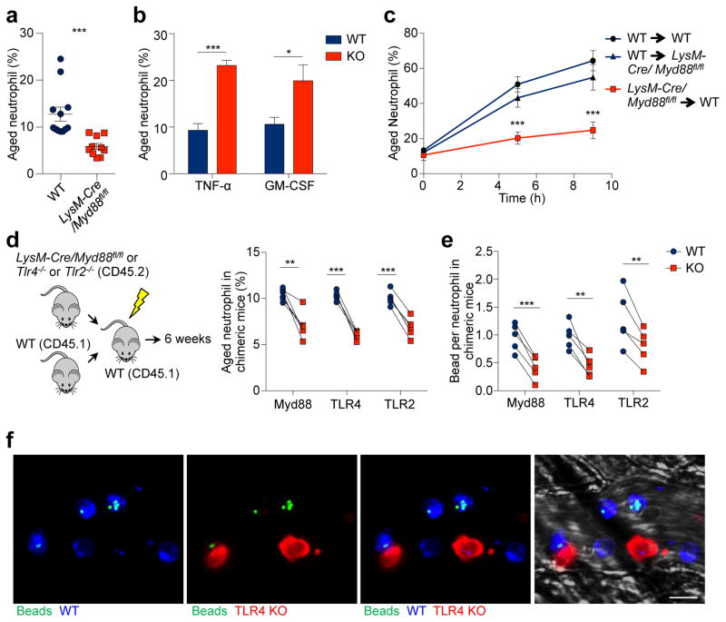
Figure 3. Microbiota-driven neutrophil ageing is mediated by neutrophil TLRs and Myd88 signalling.
a, Percentages of aged neutrophils in WT and LysM-cre/Myd88fl/fl mice, as analysed by flow cytometry (n = 12,10 mice). b, Percentages of aged neutrophils in WT and Tnf−/− or Csf2−/− mice (n = 5 mice). c, Ageing kinetics of donor neutrophils after adoptive transfer from either WT or LysM-cre/Myd88fl/fl mice into WT or LysM-cre/Myd88fl/fl recipients (n = 6 mice). d, Percentages of the aged subset in WT and LysM-cre/Myd88fl/fl, Tlr4−/− or Tlr2−/− neutrophils in chimeric mice (n = 5 mice). e, MFIM analysis of Mac-1 activation on WT and LysM-cre/Myd88fl/fl, Tlr4−/− or Tlr2−/− neutrophils in chimeric mice (n = 5 mice). f, Representative images showing WT (CD45.1+, blue) and Tlr4−/− (CD45.2+, red) neutrophils and beads (green) captured. Scale bar, 10 μm. Error bars, mean ± s.e.m. * P < 0.05, ** P < 0.01, *** P < 0.001, data representing ≥2 independent experiments analysed with unpaired Student’s t-test (a–c) or paired Student’s t-test (d–e).

