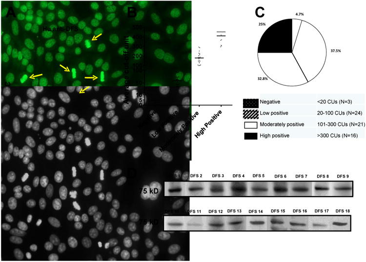
Figure 1. Detection of anti-DFS70/LEDGFp75 autoantibodies in human sera.

A) Representative human anti-DFS serum displaying the characteristic dense fine speckled nuclear pattern (FITC) in HEp-2-ANA slides. Yellow arrows point to the staining of mitotic chromosomes. Corresponding DAPI staining is shown in black and white for better visualization of chromatin. B) Scatter plot of sera with suspected anti-DFS autoantibodies that were tested using the DFS70-QuantaFlash chemiluminescent immunoassay (DFS70-CIA). C) Pie chart of data from the scatter plot. D) Blots showing immunoreactivity of selected anti-DFS sera against a 75 kD protein in whole PC3 cell lysates.
