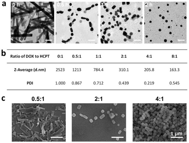
Figure 3.
(a) HD NP formation process from nanorods to nanospheres characterized by TEM at different time points (0, 0.5, 1, and 2 h). The scale bar is 500 nm. (b) Particle size distribution and polydispersity index (PDI) of particles generated from different molar ratios of DOX to HCPT (0:1, 0.5:1, 1:1, 2:1, 4:1, and 8:1). (c) SEM images of HCPT/DOX particles assembled from different molar ratios of DOX to HCPT (0.5:1, 2:1, and 4:1). The scale bar is 1 μm. When the ratio of DOX to HCPT is low, HCPT nanorods tend to form, but when the ratio of DOX to HCPT is high, spherical HD NPs are formed.

