
FIGURE 1.
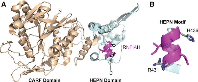
Ribbon diagram of the Pfu Csx1 monomer (PDB 4EOG). (A) (Wheat) The N-terminal modified Rossmannoid fold/CARF domain. (Pale cyan) The C-terminal winged-helix-like domain/HEPN domain. (Magenta) The highly conserved HEPN RxxxxH motif with predicted catalytic residues highlighted in black and middle residues highlighted in magenta. The dashed line represents 17 residues with missing electron density. (B) Isolated HEPN RxxxxH motif with predicted catalytic residues annotated.