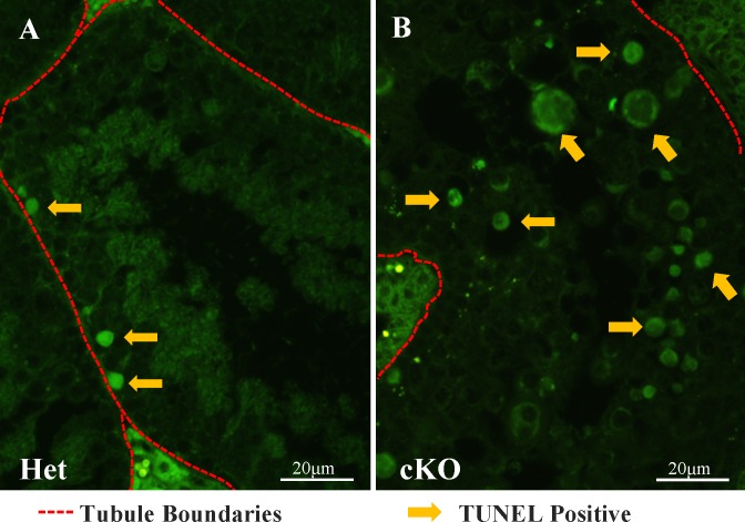
FIG. 5.
Increased TUNEL-positive cells in Cdk1 cKO testes. A and B) TUNEL staining of testis tubule sections reveals an increased number of apoptotic cells in the luminal half of the seminiferous epithelium in the cKO (B) compared to HET mice (A). Arrows indicate some TUNEL-positive cells; red hashed lines indicate tubule basal surface/perimeter. Bars = 20 μm.

