Fig. 5.
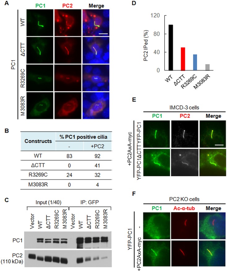
The promoting effect of PC2 on the ciliary trafficking of PC1 is dependent on its interaction with PC1 but not its CTS. (A) Ciliary localization of wild-type PC1 (WT), YFP–PC1ΔCTT, YFP–PC1-R3269C and YFP–PC1-M3083R in the presence of recombinant PC2 in IMCD-3 cells. PC1 was stained with an antibody against GFP (green) and PC2 was stained with an antibody against Myc (red). Representative images of each construct to function are shown. Scale bar: 10 µm. (B) The percentage of ciliary PC1 in the absence and presence of recombinant PC2 was scored. At least 50 ciliated, YFP–PC1-transfected cells from multiple experiments were counted for the presence of PC1 signal on cilia under each condition. (C) PC2 interaction with YFP–PC1, YFP–PC1ΔCTT, YFP–PC1-R3269C and YFP–PC1-M3083R by co-immunoprecipitation (IP). (D) Percentage of PC2 that was co-immunoprecipitated (IPed) with PC1 or its mutants. The experiment was carried out at least two times; the blot shown in Fig. 5C was used for quantification. (E) The CTS mutant PC2-AxA promotes the ciliary trafficking of both YFP–PC1 and YFP–PC1ΔCTT. YFP–PC1 and YFP–PC1ΔCTT together with PC2-AxA–Myc was transfected into IMCD-3 cells and immunofluorescence staining was performed. PC1 was detected with an antibody against GFP and PC2 was detected with an antibody against Myc. The promoting effect of PC2-AxA–Myc on YFP–PC1 and YFP–PC1ΔCTT is comparable with the wild-type PC2–Myc as described in A. Scale bar: 10 μm. (F) The CTS mutant PC2-AxA is able to rescue the ciliary trafficking defect of wild-type YFP–PC1 in PC2-KO cells. YFP–PC1 was co-transfected with or without PC2-AxA into PC2-KO cells. PC1 was detected with an anti-GFP antibody and the cilium was labeled with anti-acetylated-α-tubulin antibody (Ac-α-tub). Representative images from at least three independent experiments are shown.

