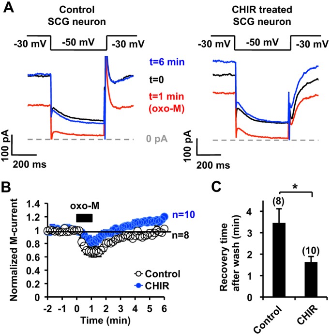
Fig. 7.

Effect of GSK3 inhibition on muscarinic suppression of the M-current. (A) Representative M-current traces from control (left) and 5 µM CHIR99021 (CHIR)-treated (right) rat SCG neurons showing suppression and recovery after treatment with 0.1 µM oxo-M. The data at the indicated time points are taken from the graph shown in B. (B) Summary time courses of oxo-M-induced M-current suppression from control and CHIR99021-treated SCG neurons, showing that the duration of oxo-M induced M-current suppression is shorter in CHIR99021-treated neurons. The black box indicates the presence of 0.1 µM oxo-M. The horizontal line indicates the baseline level of 1. (C) Quantification of the time required to recover to the control M-current amplitude after washout. *P<0.05. Error bars show s.e.m. n values are given in brackets on the graphs.
