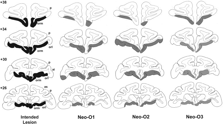
Figure 1.
Intended lesions of the orbital frontal cortex (shown in dark gray, left column) and reconstruction of the actual damage (in light gray) for the 3 cases in Group O onto standard coronal sections of a normal monkey brain. The numerals on the left of the coronal sections indicate the distance in millimeters from the interaural plane. ai, inferior arcuate sulcus; as, superior arcuate sulcus; orl, lateral orbital sulcus; orm, medial orbital sulcus; p, principal sulcus.

