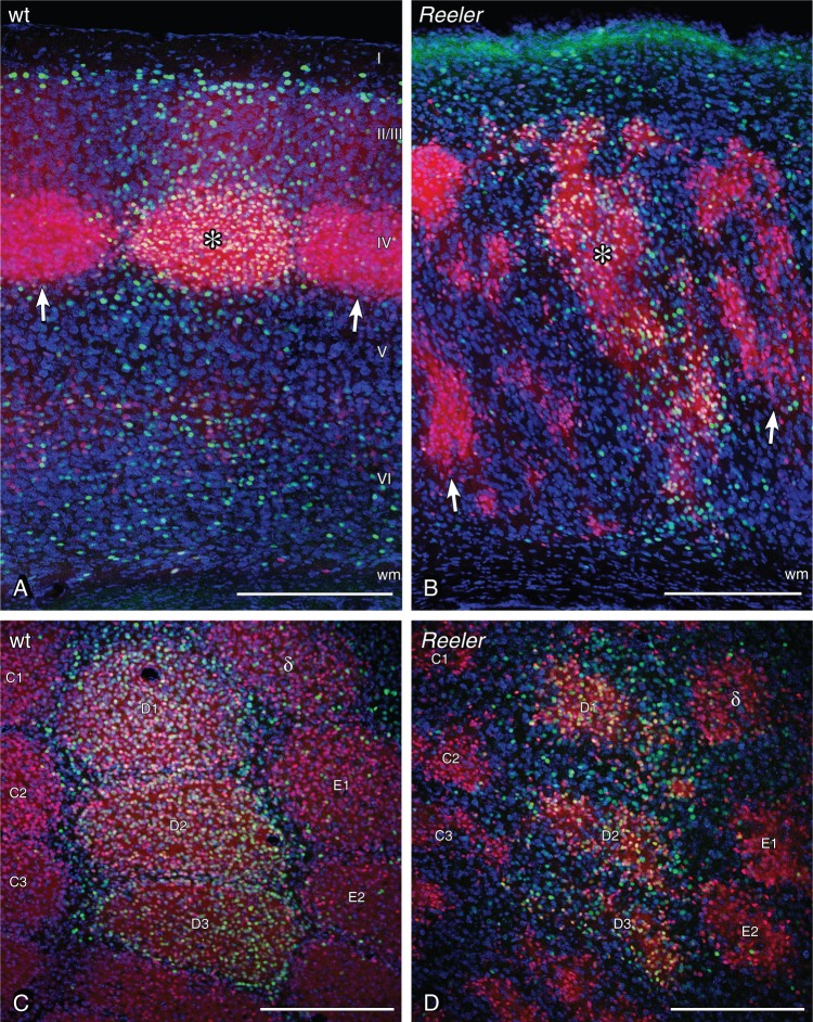
Figure 2.
Neuronal activation in the barrel-related column of LIVtdTomato wild-type and reeler mice after tactile exploration of a novel, enriched environment. The whisker corresponding to stimulated columns were used during exploration, the whiskers corresponding to unstimulated columns were clipped before exploration. (A) Primary somatosensory (S1) cortex of wild type, sectioned in the coronal plane. The barrel as a layer IV structure becomes easily visible in the transgenic animal (red neurons). The stimulated wt column shows a substantially higher amount of c-fos-positive nuclei (green staining). The stimulated column is marked by a star within the barrel, and unstimulated columns are marked by arrows at the layer IV/V border. (B) S1 reeler cortex. Barrel equivalent patches are distributed over large parts of the cortex (red cells). However, the stimulated column again becomes visible by a higher amount of c-fos-positive nuclei. The stimulated column is marked by a star within one of the stimulated barrel equivalents, unstimulated columns are marked by arrows. Wild-type (C) and reeler (D) S1 barrel cortex sectioned in the tangential plane 400 µm below the pial surface. Barrels and barrel equivalents are labeled according to standard nomenclature. D1–D3 represent stimulated columns. Scale bars: 250 µm.

