Figure 7.
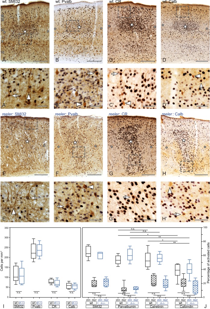
Activation of subgroups of excitatory and inhibitory wild-type and reeler neurons after tactile exploration of a novel, enriched environment. (A–H) Different subpopulation of excitatory and inhibitory neurons visualized by DAB immunostaining (brown). Nuclei of activated (c-fos-positive) neurons are revealed by DAB-Ni immunostaining (black). Stimulated columns are marked by a star within the wild-type barrel (A–D) or reeler barrel equivalent (E–H), and unstimulated columns are marked by asterisks. (A′H′) The micrographs show a higher magnification of the area delineated by the dashed frames in (A–H). Exclusively marker-labeled neurons are pointed at by arrowheads, and marker-labeled neurons that show a c-fos-positive nucleus are indicated by arrows. (I,J) Boxplots show the density of subgroups of excitatory and inhibitory neurons (I) and the strength of the activation (J). Note that stimulated columns always show a higher amount of activated neurons and that the strength of the activation is not significantly different between wild type and reeler. Bottom and top of the boxes represent the 25th and 75th percentiles, respectively; the bar in the box indicates the median. The ends of the whiskers represent the minimum or the maximum. Scale bars: A–H 200 µm; A′–H′ 40 µm.

