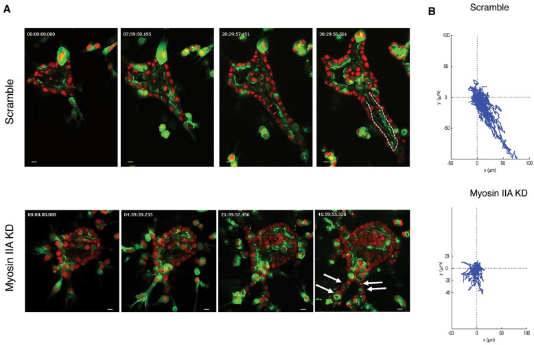
Fig. 7.
Inhibition of myosin IIA impairs directed tubular cell migration during tubulogenesis. (A) Stills of movies of tubulogenesis in control- (Scramble) (Movie 6) or myosin-IIA-depleted cysts (Movie 7). Arrows indicate multiple lumens and the dashed line outlines the lumen. Scale bars: 10 μm. (B) Migration tracks of cells are displayed as displacement plots. For each group, the trajectories of 30 to 40 cells at 30 min intervals over 24 h are presented.

