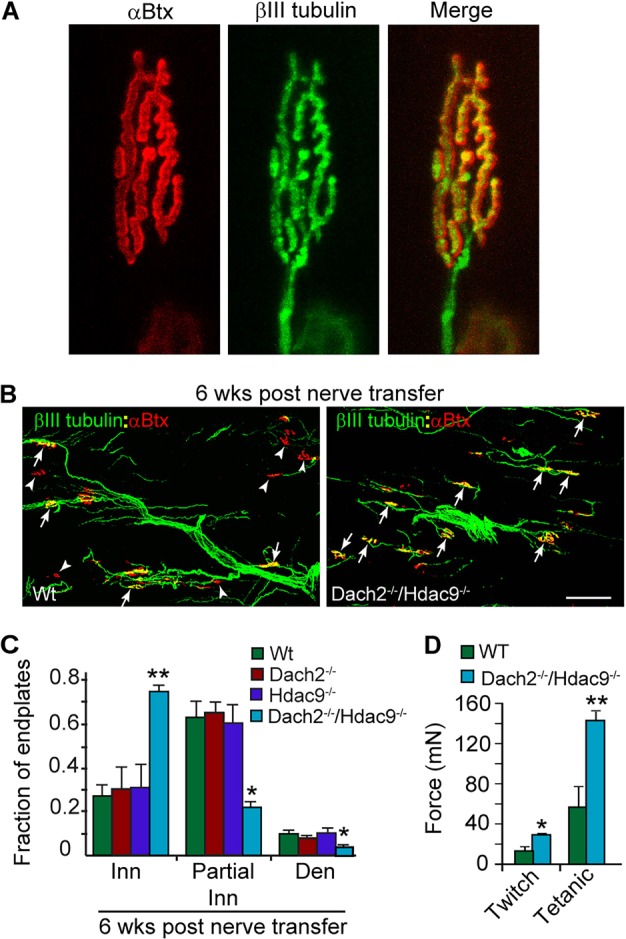
Fig. 2.

Dach2 and Hdac9 inhibit reinnervation of endplates in denervated soleus muscle following nerve transfer. (A) βIII tubulin staining of motor nerve terminals overlaps precisely with αBtx stained endplates. (B) Representative images and (C) quantification of reinnervated endplates at 6 weeks post nerve transfer. βIII tubulin+ regenerating tibial nerve branches are green and αBtx+ endplates are red. Arrows point to fully innervated endplates. Arrowheads point to denervated endplates. Scale bar: 50 µm. Error bars are s.e.m.; n=4 for Wt and Dach2−/−; n=3 for Hdac9−/− and Dach2−/−/Hdac9−/−. *P<0.05; **P<0.01 relative to Wt. (D) Soleus force measurements following indirect stimulation through the transplanted tibial nerve. Error bars are s.e.m.; n=6 for Wt and n=5 for Dach2−/−/Hdac9−/−. *P<0.05; **P<0.01 relative to Wt. Inn, innervated; Den, denervated.
