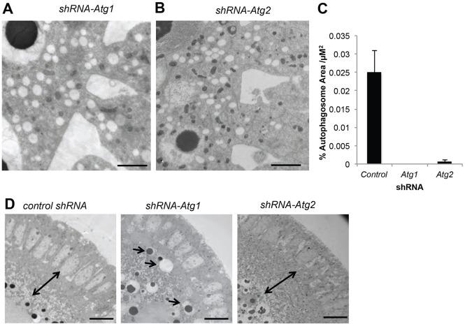
Fig. 5.
Atg1 is necessary for autophagy after cellularization. (A-C) EM of stage 7 shRNA-Atg1 (A) and shRNA-Atg2 (B) embryos, and quantification of the percent autophagosomes per µm2 in shRNA-control versus shRNA-Atg1 and shRNA-Atg2 stage 7 embryos (C) reveals a drastic reduction in formation of autophagosomes at cellularization in shRNA-Atg1 and shRNA-Atg2 embryos. Data are represented as the s.d. of three biological replicates. (D) Stage 5 embryos in shRNA-control, shRNA-Atg1 and shRNA-Atg2 embryos show a layer of lipids and mitochondria forming between nuclei and yolk in control embryos and shRNA-Atg2 embryos (double-headed arrow) that is not present in shRNA-Atg1 embryos. Arrows indicate displaced organelles in shRNA-Atg1 embryos. Scale bars: 10 µm in D; 2 µm in A,B.

