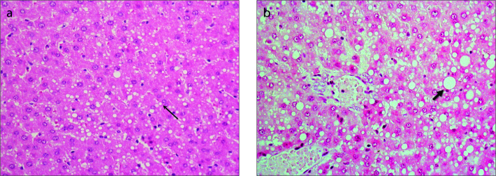
Figure 4 a, b.
Panel (a) shows histologic section of the liver and HE-staining measured 24 hours after the second ethanol intoxication with about 30% fatty degeneration including microvesicular (thin arrow) and macrovesicular fat droplets. Panel (b) shows approximately 50% mainly macrovesicular fatty degeneration of hepatocytes (thick arrow) with leukocyte demarcated single cell necrosis.

