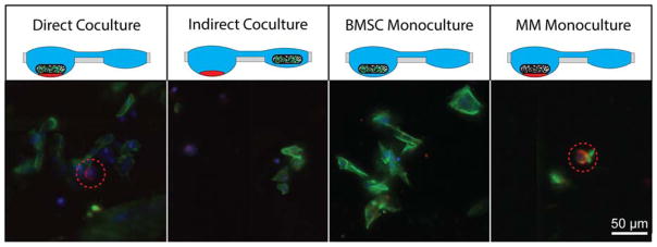
Figure 6.
Coculture and migration of MM.1S cells into polymeric bone-like scaffold. Fluorescent images of polymeric bone-like scaffolds following either direct coculture (BMSC-seeded scaffold with MM.1S cluster - same well), indirect coculture (BMSC-seeded scaffold with MM.1S - separate wells), BMSC monoculture (BMSC-seeded scaffold only), or multiple myeloma monoculture (MM.1S cultured with blank scaffold). BMSCs were identified by presence nuclear stain (blue) with actin (green), while MM.1S cells were identified by nuclear stain, actin, and CD138 ring (red) and highlighted with a red dotted circle.

