
Fig. 1.
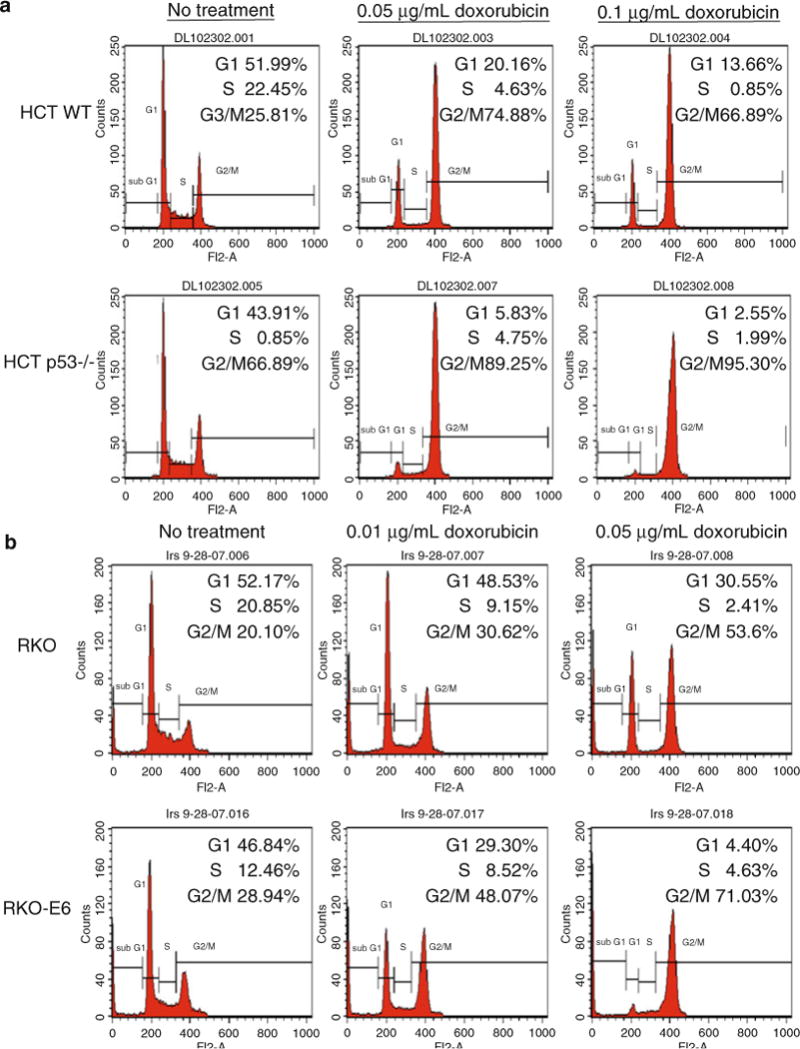
Wild-type p53 cells maintain a G1 and G2/M arrest after treatment with doxorubicin, whereas p53−/− cells primarily arrest in G2/M. (a) HCT 116 WT and p53−/− colorectal carcinoma cells were treated with 0.05 and 0.1 μg/ml doxorubicin for 24 h before harvesting for flow analysis. (b) RKO (wild-type p53) and RKO E6 (p53 is targeted for degradation by HPV E6) colorectal carcinoma cells were treated with 0.01 and 0.05 μg/ml doxorubicin for 24 h before harvesting for flow analysis. The analysis cell profiles are shown for untreated and treated cells. The percentage of cells in G1, S, and G2/M are indicated in the upper right hand corner of each histogram.