
Fig. 2.
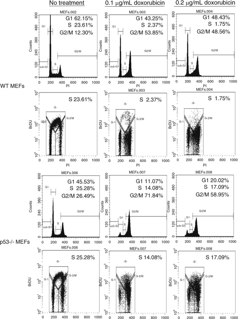
Treatment with doxorubicin in WT p53 mouse embryonic fibroblasts (MEFs) results in a significant reduction of S phase cells. Wild-type and p53−/− MEFs were treated with 0.1 and 0.2 μg/ml doxorubicin for 24 h prior to being pulsed with 10 μM BrdU for 4 h. Following antibody treatment and propidium iodide staining the cells were analyzed by dual parameter flow cytometry. Both single and dual parameter histograms are shown for each condition. The percentage of cells in G1, S, and G2/M are indicated in the upper right hand corner of each histogram.