
Fig. 3.
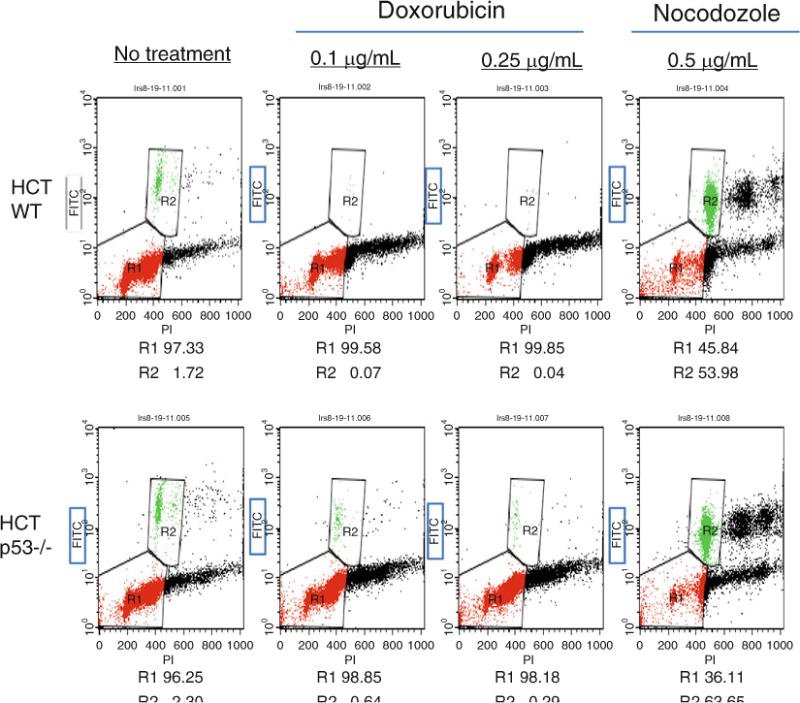
Phosphorylation of histone H3 is diminished after DNA damage with doxorubicin in WT p53 cells. HCT 116 p53 WT and −/− cells were treated with 0.1 and 0.25 μg/ml doxorubicin or 0.5 μg/ml nocodozole for 24 h and stained for Ser10-phosphorylated histone H3 and DNA content (propidium iodide). The dual parameter flow analysis is shown. Phosphorylated histone H3 positive cells are boxed and denoted as R2, whereas cells in G1 and G2 are grouped in R1.