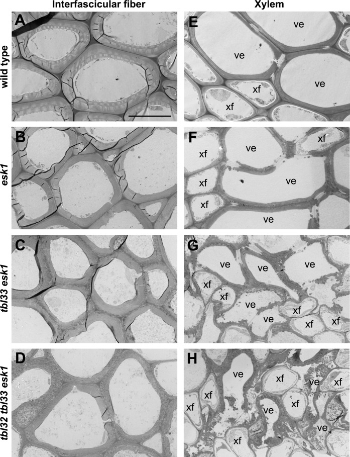
Fig 5. Transmission electron micrographs of interfascicular fibers and xylem in the wild type and various mutants.
The bottom internodes of stems of 8-week-old wild-type and esk1 plants, 12-week-old tbl33 esk1 plants, and 16-week-old tbl32 tbl33 esk1 plants were sectioned for visualization of walls of interfascicular fibers and xylem vessels. (A) to (D) Cross sections of interfascicular fibers showing defective secondary wall thickening in esk1 (B), tbl33 esk1 (C), and tbl32 tbl33 esk1 (D) compared with the wild type (A). (E) to (H) Cross sections of xylem cells showing various degrees of deformation in vessels in esk1 (F), tbl33 esk1 (G), and tbl32 tbl33 esk1 (H) compared with the wild type (E). ve, vessel; xf, xylary fiber. Bar in (A) = 10.5 μm for (A) to (H).

