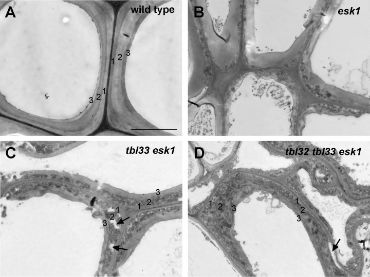
Fig 6. Effects of reduced acetylation of xylan on secondary wall structure in xylem vessels of esk1 (B), tbl33 esk1 (C) and tbl32 tbl33 esk1 (D) compared with the wild type (A).
Ultrathin stem sections were stained with lead citrate and uranyl acetate and visualized for vessel secondary wall structure under a transmission electron microscope. The numbers 1, 2 and 3 marked on vessel secondary walls denote the S1, S2 and S3 layers, respectively. Note the drastic alteration in the staining pattern of the S2 layer as well as disintegrated walls in the S2 layer (arrows) in tbl33 esk1 (C) and tbl32 tbl33 esk1 (D). Bar in (A) = 3 μm for (A) to (D).

