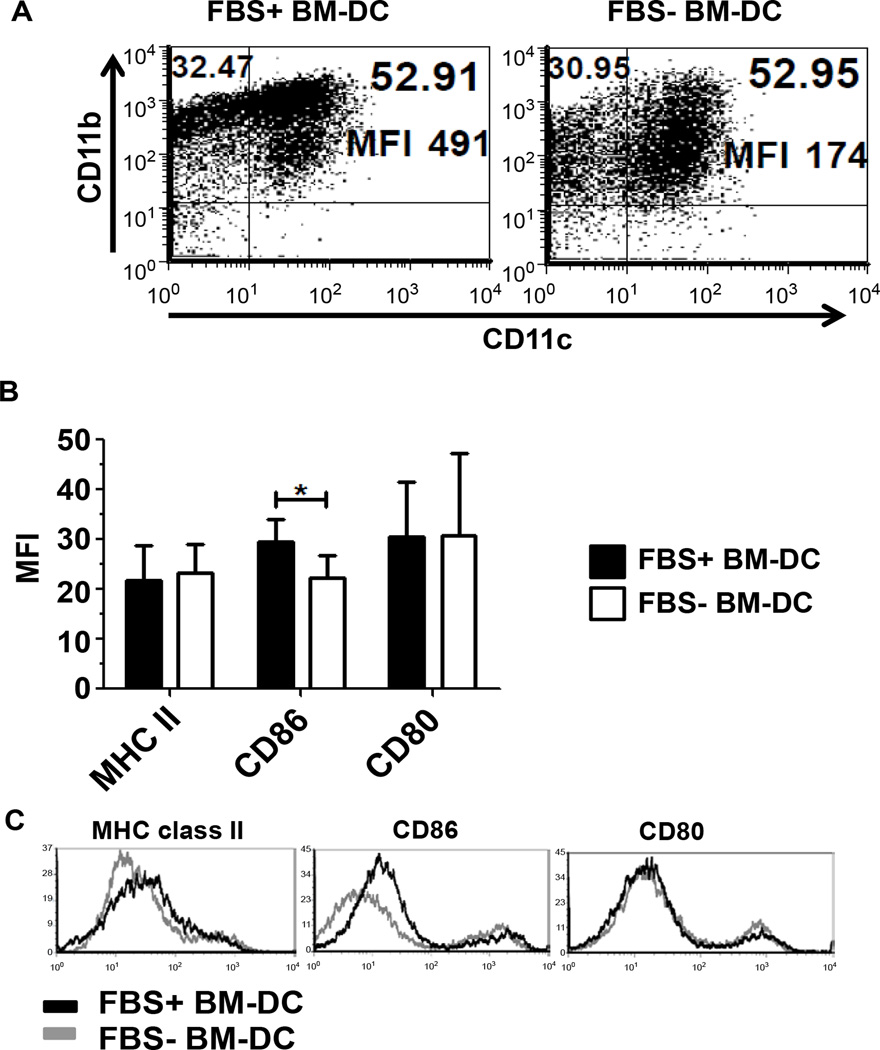
Figure 1.
Flow cytometric profile of GM-CSF and IL-4 conditioned BM-DC. BM-DC cultured in FBS− and FBS+ conditioned media were collected on Day 5 of culture and their phenotype assessed. A. The expression of CD11c and CD11b on BM-DC was assessed. B. The Mean Fluorescence Intensity (MFI) of co-stimulatory molecules CD86 and CD80, and MHC class II was assessed in three independent experiments (n=3). C. Representative histograms of the indicated markers gated on CD11c+ cells.

