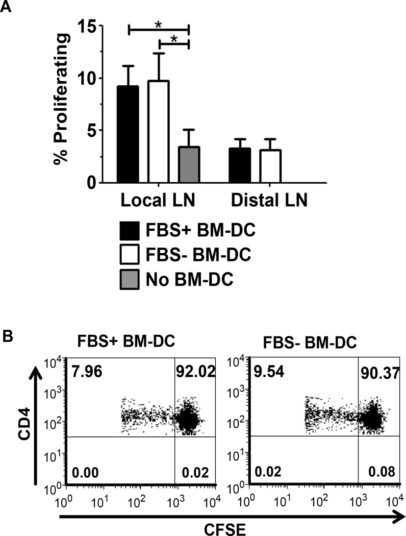
Figure 4.
In vivo BM-DC-induced CD4+ T cell proliferation. Recipient wt NOD females were subcutaneously treated with FBS+ or FBS− BM-DC pulsed with the GAD217-238 peptide or no BM-DC. 20×106 CFSE labeled splenocytes from wt NOD were simultaneously injected intravenously. On day 5 post injection, the popliteal lymph node (Local) and axial lymph node (Distal) were harvested and proliferation of CD4+ CFSE+ T cells was assessed. A. Results are shown from FBS+ BM-DC (n=5), FBS− BM-DC (n=5) and No BM-DC (n=4). B. Representative dot plots from the Local lymph nodes of FBS+ and FBS− BM-DC treated mice.

