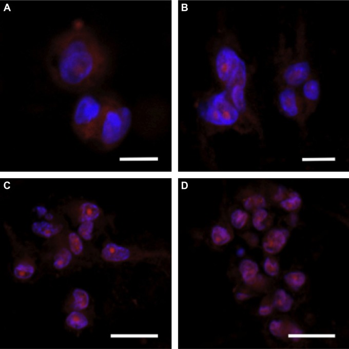
Figure 8.
Breast cancer cells cultured on collagen I hydrogels. MDA-MB-231 cells were cultured in collagen I hydrogels for one, three, five, and seven days (A–D, respectively), exhibiting the typical cell–matrix and cell–cell interactions observed in vivo. Cells developed an elongated morphology over seven days with visible processes, demonstrating cell–matrix interactions. As the cells began to proliferate, they aggregated into 3D clusters, demonstrating cell–cell interactions. Scale bars are (A, B) 10 μm and (C, D) 20 μm. Reused from Szot CS et al,71 with permission from Elsevier.

