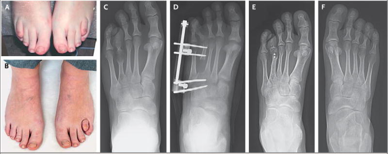
Figure 1. Images of the Toes before and after Surgery.

The patient’s shortened fourth toes before distraction osteogenesis are shown in Panel A. Panel B shows the toes after the procedure; the right toe appears normal, whereas the left toe is not completely corrected, because of an infection that required removal of the external fixators. Radiographs of the shortened left fourth metatarsal before surgical intervention are shown in Panel C. Panel D shows the left foot after osteotomy, with external fixation in place and overcorrection of the metatarsal. Panel E shows the foot after removal of the external fixation device and shortening osteotomy to relocate the joint, leaving only two screws in place. Panel F shows a normal-appearing right fourth metatarsal 9 years after distraction osteogenesis.
