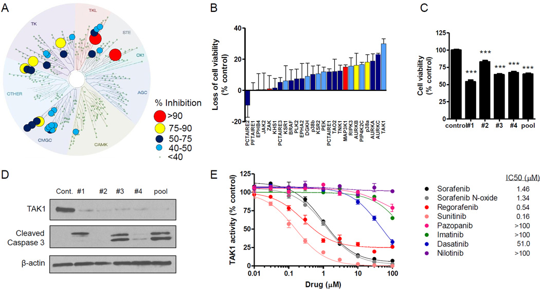
Fig. 3. TAK1 is targeted by sorafenib in human primary keratinocytes (HEKa), leading to cell death.
(A) Kinase tree illustration showing the sorafenib (10 µM) kinase target signature in HEKa using a KiNativ in situ kinase assay. (B) HEKa were transfected with siRNA (25 nM) against the top 27 kinases identified a KiNativ in situ assay, and cell viability was measured 72 h later using CellTiter-Glo (3 experiments, n = 3). (C and D) HEKa were transfected with TAK1-targeted siRNA (25 nM, individual or pooled), and 72 h later (C) cell viability was measured using CellTiter-Glo (2 experiments, n = 16) and (D) Western blot analysis was performed using the indicated antibodies. (E) Inhibition of in vitro TAK1 kinase activity by various kinase inhibitors as determined using the Millipore KinaseProfiler assay (n = 2). Data represent the mean ± SEM (***, P<0.001).

