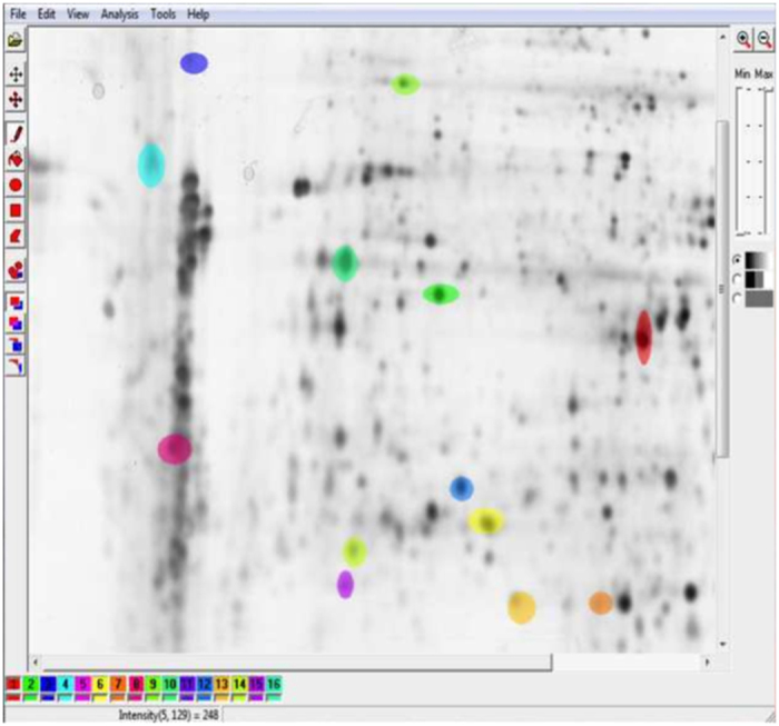
Figure 1. Two-dimensional gel electrophoresis image.

Generated using human dermal fibroblasts in order to study the effect of a plant extract on the protein expression of IBR3. 1024 × 1024 8-bit image. 2D protein separation were visualized by silver staining using standard protocols. From the dataset of G.-Z. Yang and co-workers44. Spots manually segmented using Mazda software, first step in the image analysis pipeline (dataset generation).
