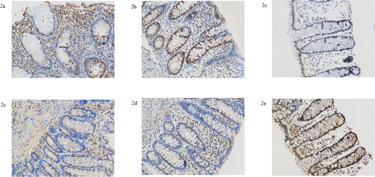
Fig 2. Representative Sigmoid Colon specimens of UC patients and patients with benign colonic polyps.
Expression of Lewis a antigen (a-c) and Lewis b antigen (d-f) in the sigmiod specimens of inflammatory and adjacent non-inflammatory tissue of UC patients and normal controls. Samples a, b, d, e were derived from patient 5, described in Table 6. Samples c and f were derived from normal tissue of patients with benign colonic polyps. Immunohistochemical staining indicated increased expression of Lewis a antigen in the cryptic epithelium in inflammatory lesions from UC patients and normal mucosa from patients with benign colonic polyps (see arrows in a-c). Expression in the epithelium did not differ dramatically between UC patients and patients with benign colonic polyps. Expression of Lewis b antigen did not differ dramatically between the three groups (d–f).

