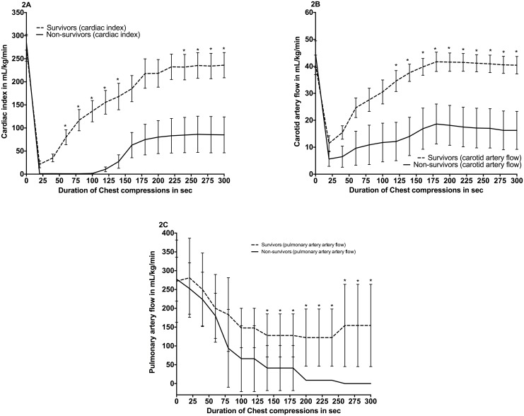
Fig 3. Cardiac index (2A), carotid artery flow (2B), pulmonary artery flow (2C) for survivors vs. non-survivors.
Baseline (at “0”, PPV until “30sec”, and CPR thereafter). Data are presented in mean (middle of line) with standard deviation (error bars), (* indicates p<0.05 survivors vs. non-survivors).

