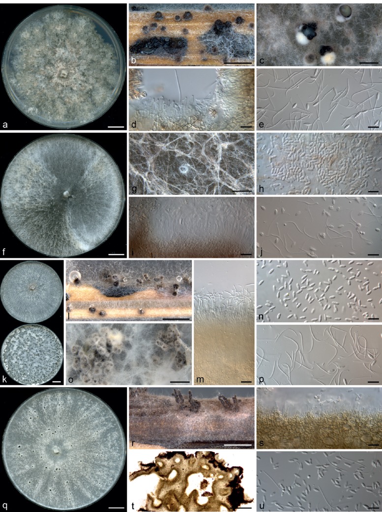
Fig. 3.
Diaporthe spp. — a–e: Diaporthe macintoshii (ex-type BRIP 55064a) after 4 wk. a. Culture on PDA; b. pycnidia on sterilised wheat straw; c. pycnidia on OMA; d. conidiophores; e. alpha conidia and beta conidia. — f–j: Diaporthe masirevicii (ex-type BRIP 57892a) after 4 wk. f. Culture on PDA; g. conidiomatum on OMA; h. alpha conidia; i. conidiophores; j. alpha conidia and beta conidia. — k–p: Diaporthe middletonii (ex-type BRIP 54884e) after 4 wk. k. Culture on PDA (top) and OMA (bottom); l. pycnidia on sterilised wheat straw; m. conidiophores; n. alpha conidia; o. pycnidia on OMA; p. beta conidia. — q–u: Diaporthe miriciae (ex-type BRIP 54736j) after 4 wk. q. Culture on PDA; r. conidiomata on sterilised wheat straw; s. conidiophores; t. section across conidiomatum; u. alpha and beta conidia. — Scale bars: a, f, k, q = 1 cm; b, c, g, l, o, r = 1 mm; d, e, h–j, m, n, p, s, u = 10 μm; t = 100 μm.

