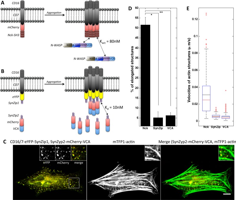
FIGURE 3:
Allowing VCA turnover in membrane clusters does not result in formation of comet tails. (A) Schematic of CD16/7-mCherry-Nck aggregation and binding by endogenous N-WASP protein. (B) Schematic of VCA clusters with turnover through SynZip-mediated interaction. CD16/7-eYFP-SynZip1 aggregation results in recruitment of cytosolic SynZip2-mCherry-VCA fusion proteins. (C) Actin recruitment and shape of actin structures induced by clustering of SynZip2-mCherry-VCA through interaction with CD16/7-eYFP-SynZip1. Left, merged staining of SynZip2-mCherry-VCA (red) and CD16/7-eYFP-SynZip1 (green). Insets, magnified views of CD16/7-eYFP-SynZip1 (left inset), SynZip2-mCherry-VCA (middle inset), and a composite of the two (right inset). Middle, actin staining alone. Right, merged staining of SynZip2-mCherry-VCA (red) and mTFP1-actin (green). Scale bar, 10μm. (D) Morphology analysis of actin structures induced by VCA clustered through SynZip binding. Nck SH3 clustering: three cells, 124 aggregates; VCA clustering though SynZip interaction: eight cells, 152 aggregates; VCA clustering: three cells, 233 aggregates. Error bars are mean ± SEM. *p < 0.001, **p < 0.01. (E) Velocity of actin structures induced by VCA clustered through SynZip binding. Nck SH3 clustering: three cells, 121 aggregates; VCA clustering though SynZip interaction: four cells, 174 aggregates; VCA clustering: three cells, 246 aggregates.

