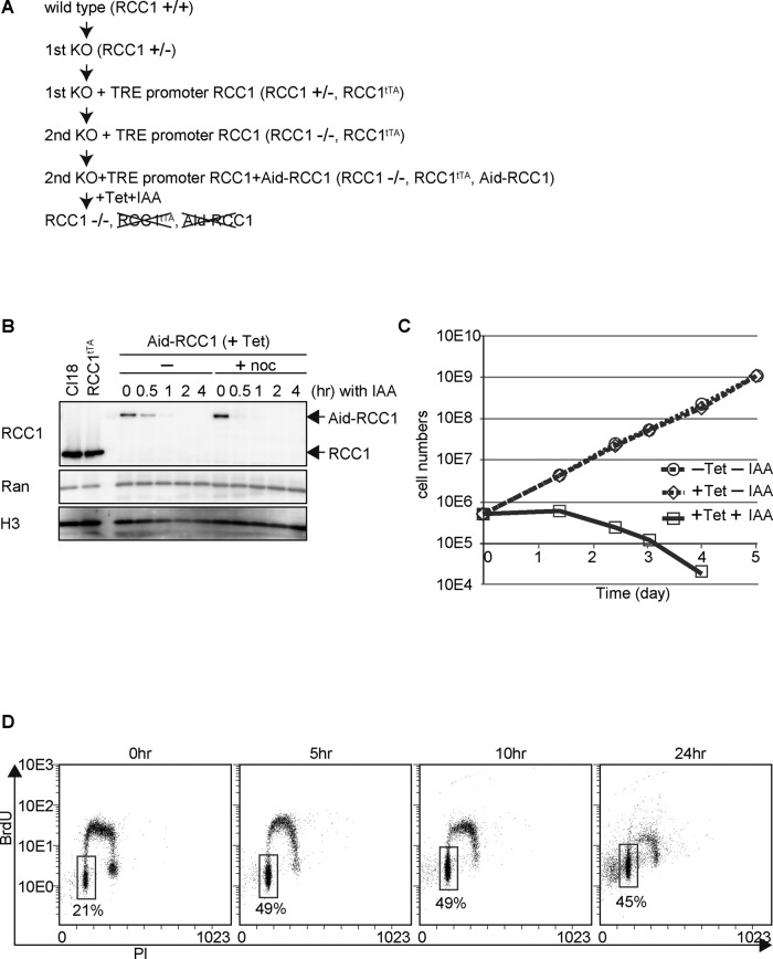
FIGURE 1:
Establishment of the Aid-based RCC1 conditional knockout cell line. (A) Scheme of Aid-based RCC1 conditional knockout cell line generation. (B) Protein levels of Aid-RCC1 in Aid-based RCC1 conditional knockout cells after addition of IAA in the presence or absence of nocodazole. Wild-type DT40 (Cl18) cells or cells expressing RCC1 cDNA (RCC1tTA) under the control of a TET-responsive promoter were also examined. Whole-cell lysates were subjected to 5–20% SDS–PAGE, followed by Western blot analysis with affinity-purified polyclonal anti-RCC1 antibody. Anti-Ran was used as a negative control, and anti–histone H3 was used as the loading control. (C) Proliferation of Aid-based RCC1 conditional knockout cells after addition of TET and IAA. Live cells were counted after trypan blue staining. (D) Cell cycle distribution after depletion of RCC1. Aid-based RCC1 conditional knockout cells were maintained in the presence of TET at a final concentration of 2 μg/ml, and samples were collected at the indicated times after addition of 500 μM IAA. Incorporation of propidium iodide (x-axis, linear scale) and BrdU (y-axis, log scale) was analyzed by FACS. The boxes represent the populations of G1 phase cells, and the numbers indicate the percentages of G1 population.

