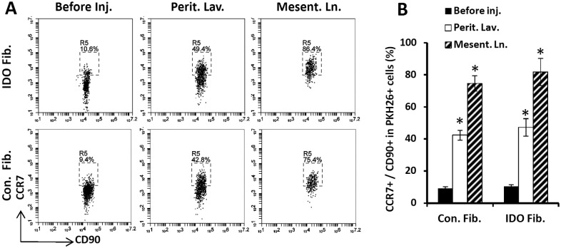
Fig 6. Expression of CC-chemokine receptor 7 (CCR7) by dermal fibroblasts.
IDO-expressing and control mouse dermal fibroblasts were examined for expression of CCR7 before and after intraperitoneal injection. To track them, fibroblasts were labeled with PKH26 red fluorescent cell membrane labeling kit. Panel A shows representative flow cytometry plots showing frequency of CCR7+ cells gated on PKH26+ CD90+ window. Upper and bottom rows show IDO-expressing and control fibroblasts data, respectively. The plots on the left side show fibroblasts before peritoneal injection (Before inj.). The middle plots and right side plots show cells that were harvested from peritoneal cavity by lavage (Perit. Lav.) or extracted from mesenteric lymph nodes (Mesent. Ln.) two weeks post IP injection, respectively. Panel B show quantification of CCR7 expression on IDO-expressing and control fibroblasts before and after IP injection in peritoneal cavity and mesentric lymph nodes. (*) denotes statistically significant difference in CCR7 expression on IDO and control fibroblasts following IP injection mice compared to the before injection level (P<0.0001, n = 5).

