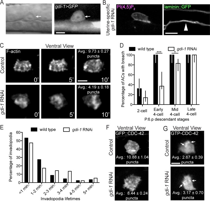
Fig 4. GDI-1 regulates invadopodia formation.
(A) A transcriptional reporter (gdi-1>GFP) revealed gdi-1 expression in the AC (arrow). (B) Uterine-specific RNAi against gdi-1 blocked AC invasion (arrowhead marks intact BM). (C) A ventral time series shows decreased invadopodia (marked with F-actin) formation in animals with reduced gdi-1 (RNAi; bottom) relative to wild type (top; overlaid text reports average number of invadopodia per timepoint from 6 wild type animals and 5 gdi-1 RNAi treated animals; p < 0.0001, Tukey’s multiple comparisons test). (D) BM breaching was delayed in gdi-1 RNAi treated animals (n ≥ 25 animals per stage, per genotype; *** p < 0.0001, * p < 0.05, Fisher’s exact test; bars represent 95% confidence intervals). (E) Animals treated with gdi-1 RNAi have longer-lived invadopodia (n = 6 wild type and 7 gdi-1 RNAi treated animals; p < 0.01, Chi-square test). (F) Ventral views showing that the wild type, punctate pattern of GFP::CDC-42 (top) was not affected by loss of gdi-1 (bottom; overlaid text reports average number of puncta from 8 wild type animals and 9 gdi-1 RNAi treated animals; p = 0.05, Student’s t-test). (G) CDC-42 was still activated normally (as visualized with the GTP-CDC-42 sensor) after loss gdi-1 (overlaid text reports average number of puncta from 18 wild type animals and 6 gdi-1 RNAi treated animals; p = 0.53, Student’s t-test). Scale bars = 5 μm.

