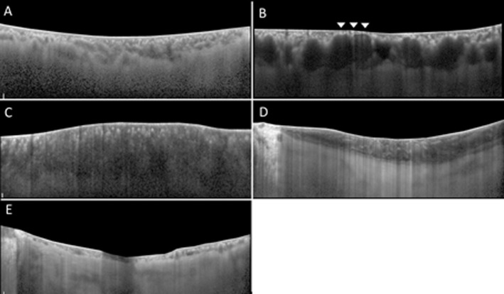
Fig 1. Representative images of the choroid.
These demonstrate 5 types of tomographic classification in current study, after elimination of retinal image above retinal pigment epithelium. Standard (S) type (Fig 1A) shows clear visualization of the inner choriocapillaris layer, medium choroidal vessel layer and large choroidal vessel layer. The choroidal vessels are irregularly distributed among the choroidal layer and its lumen heterogeneous. The chorio-scleral border is relatively distinct and well-defined. Dilated outer layer and Attenuated inner layer (DA) type (Fig 1B) shows dilation of large choroidal vessel accompanied with the invisibility of innermost layer in the focal area of choroid (arrowheads). Darkened (D) type (Fig 1C) shows diffuse homogenous hyporeflectivity and, markedly decreased visibility of the outer choroidal layer. Marbled (M) type (Fig 1D) shows slightly dim choroidal reflectivity with obscured vessel margins. Multiple amorphous hyperreflective materials are dispersed within layers. In Pauci-Vascular (PV) type (Fig 1E), there are a lack of large choroidal vessels with choroidal thickness less than 100 um in the fovea and extrafoveal areas.

