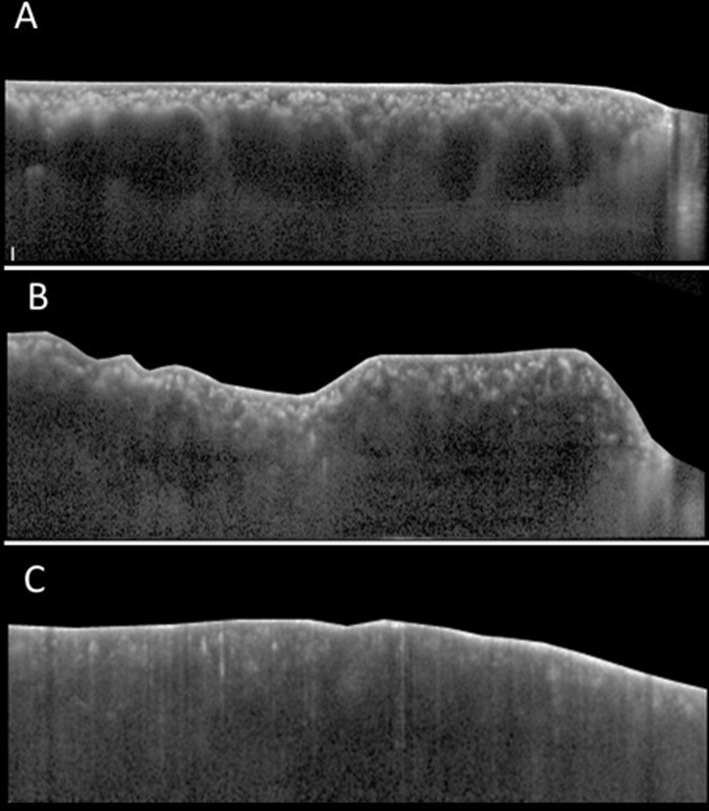
Fig 2. Additional tomographic characteristics of the choroid.
Choroidal vessel dilation (Fig 2A): the presence of 3 consecutive vessel lumens with a diameter width of at least 200㎛ in the large choroidal vessel layer is observed. Homogenous hyperreflectivity consistent from the RPE line to the posterior border of the sclera with a constant width of at least 200㎛ is observed (arrows). Convoluted choroid (Fig 2B): the innermost choroidal layer together with the RPE line has a wavy, flexuous appearance. Scleral invisibility (Fig 2C): the chorio-scleral border beneath the choroidal layer is invisible.

