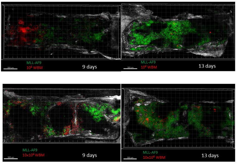
Figure 6. Sternal bone marrow confocal combined with two-photon imaging demonstration of leukemic and normal HSCs foci.
Representative micrographs of sternal marrow sections from mice transplanted with normal WBM (marked by dsRED) only compared with fixed amount of MLL-AF9 cells (marked by GFP) along with different LSKs doses. Scale bars indicate 300 μm. Two mice were imaged for each time point in each group, in two independent experiments.

