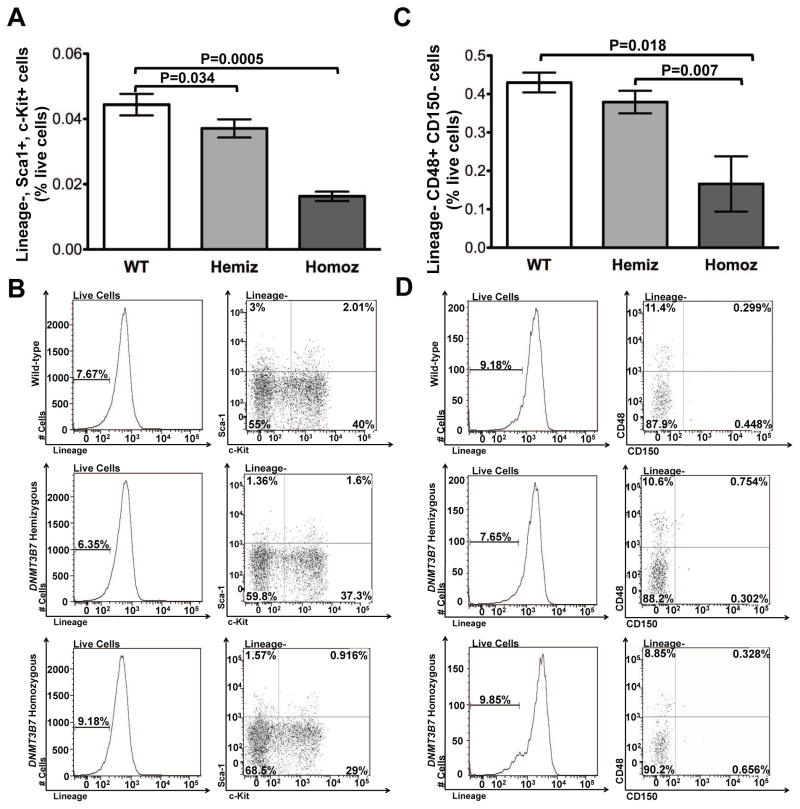
Figure 1. DNMT3B7 homozygous E14.5 embryos have fewer numbers of HSPCs.
(A) Lineage (FITC)-, Sca-1 (PE)+, c-Kit (APC)+ cells were stained in fetal livers isolated from E14.5 embryos that were Wild-type (WT: white), hemizygous DNMT3B7 transgenic (Hemiz: light grey) and homozygous DNMT3B7 transgenic (Homoz: dark grey). Average percentages from n ≥ 5 per group ± SEM are plotted. Two-tailed Student’s t-test was used to determine statistical significance. (B) Representative plots analyzed using FlowJo to quantify the relative levels of the analyzed populations. (C) Lineage (FITC)-, CD48 (PE)+, CD150 (APC)+ cells were stained in fetal livers isolated from E14.5 embryos that were Wild-type (WT: white), hemizygous DNMT3B7 transgenic (Hemiz.: light grey) and homozygous DNMT3B7 transgenic (Homoz.: dark grey). Average percentages from n ≥ 4 per group ± SEM are plotted. Two-tailed Student’s t-test was used to determine statistical significance. (D) Representative plots analyzed using FlowJo to quantify the relative levels of the analyzed populations.

